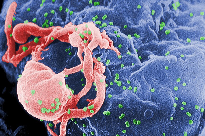
चित्र:HIV-budding-Color.jpg

इस पूर्वावलोकन का आकार: 800 × 531 पिक्सेल। दूसरे रेसोल्यूशन्स: 320 × 213 पिक्सेल | 640 × 425 पिक्सेल | 1,024 × 680 पिक्सेल | 1,280 × 850 पिक्सेल | 2,967 × 1,971 पिक्सेल।
मूल चित्र ((2,967 × 1,971 पिक्सेल, फ़ाइल का आकार: 3.92 MB, MIME प्रकार: image/jpeg))
चित्र का इतिहास
फ़ाइलका पुराना अवतरण देखने के लिये दिनांक/समय पर क्लिक करें।
| दिनांक/समय | थंबनेल | आकार | सदस्य | प्रतिक्रिया | |
|---|---|---|---|---|---|
| वर्तमान | 00:16, 20 अप्रैल 2008 |  | 2,967 × 1,971 (3.92 MB) | Optigan13 | {{Information |Description={{en|Scanning electron micrograph of HIV-1 budding from cultured lymphocyte. See PHIL 1197 for a black and white view of this image. Multiple round bumps on cell surface represent sites of assembly and budding of virions.}} |Sou |
चित्र का उपयोग
निम्नलिखित पन्ने इस चित्र से जुडते हैं :
चित्र का वैश्विक उपयोग
इस चित्र का उपयोग इन दूसरे विकियों में किया जाता है:
- ar.wiki.x.io पर उपयोग
- arz.wiki.x.io पर उपयोग
- ast.wiki.x.io पर उपयोग
- as.wiki.x.io पर उपयोग
- azb.wiki.x.io पर उपयोग
- az.wiki.x.io पर उपयोग
- be-tarask.wiki.x.io पर उपयोग
- bg.wiki.x.io पर उपयोग
- bn.wiki.x.io पर उपयोग
- ca.wiki.x.io पर उपयोग
- ca.wikinews.org पर उपयोग
- ckb.wiki.x.io पर उपयोग
- cs.wiki.x.io पर उपयोग
- Wikipedie:Studenti píší Wikipedii/Pokroky v imunologii I (2013/2014)
- Wikipedie:Studenti píší Wikipedii/Pokroky v imunologii I (2014/2015)
- Wikipedie:Nástěnka/Univerzita Karlova/Pokroky v imunologii (2013-2014)
- Wikipedie:Nástěnka/Univerzita Karlova/Molekulární imunologie (2014-2015)
- Wikipedie:Nástěnka/Univerzita Karlova/Pokroky v imunologii (2014-2015)
- cy.wiki.x.io पर उपयोग
- de.wiki.x.io पर उपयोग
- diq.wiki.x.io पर उपयोग
- en.wiki.x.io पर उपयोग
- en.wikibooks.org पर उपयोग
इस चित्र के वैश्विक उपयोग की अधिक जानकारी देखें।


